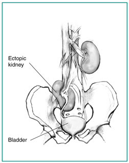
What is an ectopic kidney?
An ectopic kidney is a birth defect in which a kidney is located below, above, or on the opposite side of its usual position. About one in 900 people has an ectopic kidney.1
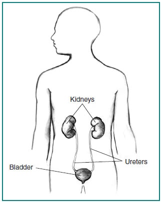
What are the kidneys and what do they do?
The kidneys are two bean-shaped organs, each about the size of a fist. They are located near the middle of the back, just below the rib cage, one on each side of the spine. Every minute, a person’s kidneys filter about 3 ounces of blood, removing wastes and extra water. The wastes and extra water make up the 1 to 2 quarts of urine a person produces each day. The urine flows to the bladder through tubes called ureters where it is stored until being released through urination.
What causes an ectopic kidney?
During fetal development, a baby’s kidneys first appear as buds inside the pelvis—the bowl-shaped bone that supports the spine and holds up the digestive, urinary, and reproductive organs—near the bladder. As the kidneys develop, they move gradually toward their usual position in the back near the rib cage. Sometimes, one of the kidneys remains in the pelvis or stops moving before it reaches its usual position. In other cases, the kidney moves higher than the usual position. Rarely does a child have two ectopic kidneys.
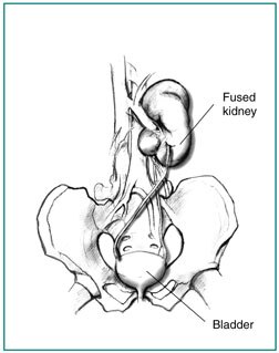
Most kidneys move toward the rib cage, but one may cross over so that both kidneys are on the same side of the body. When a crossover occurs, the two kidneys often grow together and become fused.
Factors that may lead to an ectopic kidney include
- poor development of a kidney bud
- a defect in the kidney tissue responsible for prompting the kidney to move to its usual position
- genetic abnormalities
- the mother being sick or being exposed to an agent, such as a drug or chemical, that causes birth defects
What are the symptoms of an ectopic kidney?
An ectopic kidney may not cause any symptoms and may function normally, even though it is not in its usual position. Many people have an ectopic kidney and do not discover it until they have tests done for other reasons. Sometimes, a health care provider may discover an ectopic kidney after feeling a lump in the abdomen during an examination. In other cases, an ectopic kidney may cause abdominal pain or urinary problems.
What are the possible complications of an ectopic kidney?
Possible complications of an ectopic kidney include problems with urine drainage from that kidney. Sometimes, urine can even flow backwards from the bladder to the kidney, a problem called vesicoureteral reflux (VUR). More information about VUR is provided in the NIDDK health topic, Vesicoureteral Reflux.
Abnormal urine flow and the placement of the ectopic kidney can lead to various problems:
- Infection. Normally, urine flow washes out bacteria and keeps them from growing in the kidneys and urinary tract. When a kidney is out of the usual position, urine may get trapped in the ureter or in the kidney itself. Urine that remains in the urinary tract gives bacteria the chance to grow and spread. Symptoms of a urinary tract infection include frequent or painful urination, back or abdominal pain, fever, chills, and cloudy or foul-smelling urine.
- Stones. Urinary stones form from substances found in the urine, such as calcium and oxalate. When urine remains in the urinary tract for too long, the risk that these substances will have time to form stones is increased. Symptoms of urinary stones include extreme pain in the back, side, or pelvis; blood in the urine; fever or chills; vomiting; and a burning feeling during urination.
- Kidney damage. If urine backs up all the way to the kidney, damage to the kidney can occur. As a result, the kidney can’t filter wastes and extra water from the blood. One ectopic kidney, even when it has no function, will not cause kidney failure. The other kidney can usually perform the functions of two healthy kidneys. Total kidney failure happens only in rare cases when both kidneys are damaged.
- Trauma. If the ectopic kidney is in the lower abdomen or pelvis, it may be susceptible to injury from blunt trauma. People with an ectopic kidney who want to participate in body contact sports may want to wear protective gear.
How is an ectopic kidney diagnosed?
A health care provider may use one or more of the following imaging tests to diagnose an ectopic kidney:
- Ultrasound. An ultrasound uses a device, called a transducer, that bounces safe, painless sound waves off organs to create an image of their structure. The procedure is performed in a health care provider’s office, outpatient center, or hospital by a specially trained technician, and the images are interpreted by a radiologist—a doctor who specializes in medical imaging; anesthesia is not needed. The images can show the location of the kidneys.
- Intravenous pyelogram (IVP). An IVP is an x ray of the urinary tract. A special dye, called contrast medium, is injected into a vein in the person’s arm, travels through the body to the kidneys, and makes urine visible on the x ray. The procedure is performed in a health care provider’s office, outpatient center, or hospital by an x-ray technician, and the images are interpreted by a radiologist; anesthesia is not needed. An IVP can show a blockage in the urinary tract. In children, ultrasounds are usually done instead of IVPs.
- Voiding cystourethrogram (VCUG). A VCUG is an x-ray image of the bladder and urethra taken while the bladder is full and during urination, also called voiding. The bladder and urethra are filled with contrast medium to make the structures clearly visible on the x-ray images. The x-ray machine captures images of the contrast medium while the bladder is full and when the person urinates. The procedure is performed in a health care provider’s office, outpatient center, or hospital by an x-ray technician supervised by a radiologist, who then interprets the images. Anesthesia is not needed, but sedation may be used for some people. The test can show abnormalities of the inside of the urethra and bladder and whether urine is backing up toward the kidneys during urination.
- Radionuclide scan. A radionuclide scan is an imaging technique that relies on the detection of small amounts of radiation after injection of radioactive chemicals. Because the dose of the radioactive chemicals is small, the risk of causing damage to cells is low. Special cameras and computers are used to create images of the radioactive chemicals as they pass through the kidneys. The procedure is performed in a health care provider’s office, outpatient center, or hospital by a specially trained technician, and the images are interpreted by a radiologist; anesthesia is not needed. This test can show the location of an ectopic kidney and whether the ureters are blocked.
- Magnetic resonance imaging (MRI). MRI machines use radio waves and magnets to produce detailed pictures of the body’s internal organs and soft tissues without using x rays. An MRI may include the injection of contrast medium. With most MRI machines, the person lies on a table that slides into a tunnel-shaped device that may be open ended or closed at one end; some newer machines are designed to allow the person to lie in a more open space. The procedure is performed in an outpatient center or hospital by a specially trained technician, and the images are interpreted by a radiologist; anesthesia is not needed though light sedation may be used for people with a fear of confined spaces. MRIs can show the location of the kidneys.
In addition to imaging tests, blood tests may be done to determine how well the kidneys are working. These tests are almost always normal in people with an ectopic kidney, even if it is badly damaged, because the other kidney usually has completely normal function.
How is an ectopic kidney treated?
No treatment for an ectopic kidney is needed if urinary function is normal and no blockage of the urinary tract is present.
If tests show an obstruction, surgery may be needed to correct the position of the kidney to allow for better drainage of urine. Reflux can be corrected by surgery to alter the ureter or injection of a gellike liquid into the bladder wall near the opening of the ureter.
If extensive kidney damage has occurred, surgery may be needed to remove the kidney. As long as the other kidney is working properly, losing one kidney should have no adverse health effects. More information is provided in the NIDDK health topic, Solitary Kidney.
With the right testing and treatment, if needed, an ectopic kidney should cause no serious long-term health problems.
Eating, Diet, and Nutrition
Eating, diet, and nutrition have not been shown to play a role in causing or preventing an ectopic kidney.
Points to Remember
- An ectopic kidney is a birth defect in which a kidney is located below, above, or on the opposite side of its usual position.
- Factors that may lead to an ectopic kidney include
- poor development of a kidney bud
- a defect in the kidney tissue responsible for prompting the kidney to move to its usual position
- genetic abnormalities
- the mother being sick or being exposed to an agent, such as a drug or chemical, that causes birth defects
- An ectopic kidney may not cause any symptoms and may function normally, even though it is not in its usual position.
- Possible complications of an ectopic kidney include problems with urine drainage from that kidney. Abnormal urine flow and the placement of the ectopic kidney can lead to various problems such as infection, stones, kidney damage, and injury from trauma.
- No treatment for an ectopic kidney is needed if urinary function is normal and no blockage of the urinary tract is present. Surgery or other treatment may be needed if there is an obstruction, reflux, or extensive damage to the kidney.
References
Clinical Trials
The National Institute of Diabetes and Digestive and Kidney Diseases (NIDDK) and other components of the National Institutes of Health (NIH) conduct and support research into many diseases and conditions.
What are clinical trials, and are they right for you?
Clinical trials are part of clinical research and at the heart of all medical advances. Clinical trials look at new ways to prevent, detect, or treat disease. Researchers also use clinical trials to look at other aspects of care, such as improving the quality of life for people with chronic illnesses. Find out if clinical trials are right for you .
What clinical trials are open?
Clinical trials that are currently open and are recruiting can be viewed at www.ClinicalTrials.gov.
June 2012
No comments:
Post a Comment